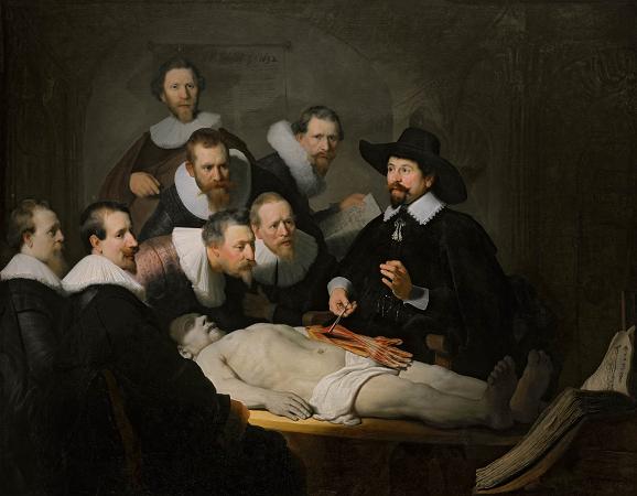
Anatomy Lesson of Nicolaes Tulp (1632). Oil on canvas. 170 x 217. The Anatomy Lesson of Dr. Nicolaes Tulp is a 1632 oil painting on canvas by Rembrandt housed in the Mauritshuis museum in The Hague, the Netherlands. The painting is regarded as one of Rembrandt's early masterpieces. In the work, Dr. Nicolaes Tulp is pictured explaining the musculature of the arm to medical professionals. Some of the spectators are various doctors who paid commissions to be included in the painting. The painting is signed in the top-left hand corner Rembrandt. f 1632. This may be the first instance of Rembrandt signing a painting with his forename as opposed to the monogramme RHL, and is thus a sign of his growing artistic confidence. The event can be dated to 31 January 1632: the Amsterdam Guild of Surgeons, of which Tulp was official City Anatomist, permitted only one public dissection a year, and the body would have to be that of an executed criminal. Anatomy lessons were a social event in the 17th century, taking place in lecture rooms that were actual theatres, with students, colleagues and the general public being permitted to attend on payment of an entrance fee. The spectators are appropriately dressed for this social occasion. It is thought that the uppermost and farthest left figures were added to the picture later. Every five to ten years, the Surgeon's Guild would commission a portrait by a leading portraitist of the period; Rembrandt was commissioned for this task when he was 26 years old, and newly arrived in Amsterdam. It was his first major commission in Amsterdam. Each of the men included in the portrait would have paid a certain amount of money to be included in the work, and the more central figures probably paid more, even twice as much. Rembrandt's anatomical portrait radically altered the conventions of the genre, by including a full length corpse in the center of the image and creating not just a portrait but a dramatic Mise-en-scone. Rembrandt's image is a fiction; in a typical anatomy lesson, the surgeon would begin by opening the chest cavity and thorax because the internal organs there decay most rapidly. One person is missing: the Preparator, whose task was to prepare the body for the lesson. In the 17th century an important scientist such as Dr. Tulp would not be involved in menial and bloody work like dissection, and such tasks would be left to others. It is for this reason that the picture shows no cutting instruments. Instead we see in the lower right corner an enormous open textbook on anatomy, possibly the 1543 De humani corporis fabrica by Andreas Vesalius. The corpse is that of the criminal Aris Kindt, who was convicted for armed robbery and sentenced to death by hanging. He was executed earlier on the same day of the scene. The face of the corpse is partially shaded, a suggestion of umbra mortis, a technique that Rembrandt was to use frequently. The French art historian Jean-Marie Clarke points out that the navel of the corpse has the shape of a capital R and connects this observation to the fact that Rembrandt worked intensively on his signatures in 1632, using three types consecutively before settling on the final, first name form in 1633. Kindt was discussed in the 1999 novel The Rings of Saturn by W. G. Sebald, and plays a significant role in Laird Hunt's 2006 novel The Exquisite. In her 2014 novel, The Anatomy Lesson author and journalist Nina Siegal tells the life story of Aris Kindt, based on documents about his criminal history that she discovered in the Amsterdam city archives. Medical specialists have commented on the accuracy of muscles and tendons painted by the 26-year-old Rembrandt. It is not known where he obtained such knowledge; it is possible that he copied the details from an anatomical textbook. However, in 2006 Dutch researchers recreated the scene with a male cadaver, revealing several discrepancies of the exposed left forearm compared to that of a real corpse. The surgically astute will notice that the origin of the exposed forearm muscles would seem to indicate that the flexor compartment originates at the lateral epicondyle, when it is, in fact, the medial epicondyle. It is the common extensor origin that originates at the lateral epicondyle. In a 2007 study, the American artist and anatomist David J. Jackowe and his colleagues demonstrated that the mysterious white cord that courses along the ulnar aspect of the cadaver's carpus and little finger, long thought to be either an ulnar nerve variant or artistic error, is most likely the tendon of an anomalous forearm muscle, the accessory abductor digiti minimi. The Anatomy Lesson of Dr. Deijman, painted by Rembrandt in 1656, was intended to be displayed in the Anatomical Hall in Amsterdam alongside The anatomy lesson of Tulp.
more...


